The Razor’s Edge of Neurooncology
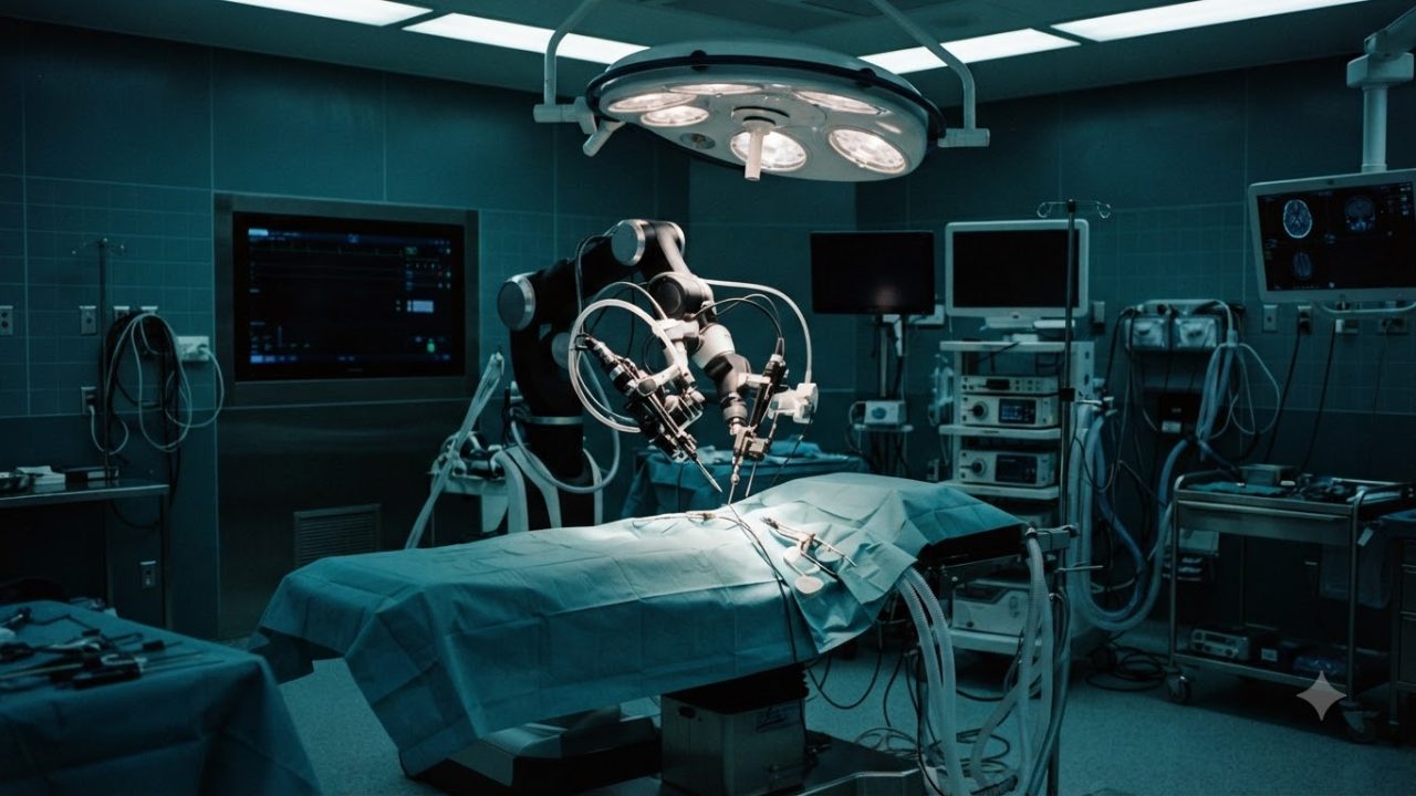
The silence in a neurosurgical theater is never truly silent. It is a heavy, pressurized atmosphere where the hum of high-end equipment serves as the metronome for a race against time and biology. At the center of this environment, a surgeon stands over the exposed brain of a patient, holding instruments that measure their impact in fractions of a millimeter. In this delicate landscape, the margins are unforgiving. A single tremor, a slight miscalculation of depth, or the misidentification of a tumor boundary against the vibrant, pulsating geography of the cerebral cortex can mean the difference between a life reclaimed and a life irrevocably altered.
For decades, the greatest anxiety for any neurosurgeon operating near the base of the skull has been the optic chiasm. This tiny, cross-shaped structure is where the nerves from both eyes converge and partially cross, and it is frequently threatened by pituitary tumors and skull-base neoplasms. To cut too deep is to court permanent blindness; to leave too much of the tumor behind is to invite a devastating recurrence. It is a razor’s edge existence that has defined the limits of human capability for generations. Yet, in London, a team of pioneering neurosurgeons has recently shattered these traditional constraints.
By successfully executing the world’s first AI-assisted brain tumor removal, they have introduced a new variable into the surgical equation—an intelligent partner capable of seeing what the human eye might overlook. This breakthrough represents more than just a technological curiosity; it is a fundamental shift in how we perceive the limitations of human anatomy and the future of invasive medicine, marking the moment when the operating room began to evolve from a purely manual theater into an augmented one.
A Historic Procedure in London
This historic milestone in London was not the result of a robotic hand taking the lead, but rather the integration of sophisticated intelligence into the surgeon’s field of vision. The operation was defined by its objective: the total excision of a complex tumor, a process historically fraught with the danger of damaging vital visual pathways. During the procedure, the surgeons relied on a fusion of classical surgical technique and real-time algorithmic processing. As the neurosurgeon navigated the cavernous spaces near the optic nerve, the AI did not replace the hand; it informed the mind.
In the past, the surgeon’s primary toolkit was limited to pre-operative magnetic resonance imaging—static, frozen in time—and their own years of training in visual pattern recognition. But tumors, especially deep-seated ones, often possess an uncanny ability to masquerade as healthy, functional tissue, blending into the surrounding architecture of the brain. The London team leveraged an active, live collaboration that allowed them to differentiate between the rogue cells of the tumor and the essential, delicate structures that preserve a patient’s sight.
By bridging this gap in real-time, the team achieved what was once considered a high-risk gamble, protecting the optic nerve while achieving a degree of resection that was previously safer to approach with extreme caution. This successful integration of human judgment and machine precision serves as the first real-world proof that the operating microscope can be transformed into a portal of deep diagnostic clarity. To understand how this was possible, one must look at the digital architecture beneath the lens. Modern intraoperative AI systems operate by transforming the surgeon’s primary perspective—the view through the oculars of a surgical microscope—into a data stream for high-resolution computer vision.
How Live AI Diagnostics Work under the Lens
This is not merely a high-definition image; it is a landscape being continuously mapped by deep learning algorithms trained on thousands of hours of surgical footage. These neural networks have essentially ‘studied’ the cellular patterns, colorations, and vascular density variations of thousands of brain tumors. In the operating theater, as the surgeon carefully dissects, the AI performs a rapid, invisible interrogation of every pixel. It identifies structural anomalies that the human eye might ignore, noting subtle changes in tissue density that distinguish a malignancy from the surrounding neurovasculature. Most importantly, this technology delivers its findings through augmented reality overlays.
As the surgeon looks into the microscope, a heads-up display projects a dynamic border over the surgical field, precisely outlining the tumor’s margins. It is as if the surgeon has been given a map that updates itself with every stroke of the dissector. This live feedback loop allows the neurosurgeon to make split-second decisions with the confidence that they are operating precisely on the boundary of the pathology. This is a dramatic departure from the way surgical diagnostics have been handled for over a century, as the surgeon is no longer forced to rely on their intuition and the stagnant reference of a film taken days or weeks prior.
Historically, the neurosurgeon’s only recourse when faced with an uncertain tissue margin was to pause the entire procedure and rely on the legacy pathway of frozen section pathology. This meant carefully extracting a small sample of tissue, handing it off to a technician, and waiting. The operating room would effectively go dark while a pathologist in a distant lab sectioned, stained, and examined the sample under a microscope to provide a diagnosis—a process that typically consumes twenty to forty minutes of critical surgical time.
The Slow, Costly Legacy of Intraoperative Pathology
Every minute spent waiting is a minute the patient remains under the heavy physiological burden of general anesthesia, which carries its own inherent risks, including post-operative confusion and increased susceptibility to infection. Furthermore, the sheer financial cost of an idle operating room, equipped with state-of-the-art technology and a full surgical staff, is staggering. When a surgeon has to stop and start repeatedly to verify if they are still within the tumor margin, the efficiency of the surgery drops, and the cost to the healthcare system spikes.
The uncertainty of the biopsy process has long been a structural limitation in neurosurgery, forcing surgeons to choose between being overly aggressive—risking damage to vital tissue—or being overly conservative, leaving behind tumor fragments that will eventually require a secondary intervention. The introduction of real-time, AI-assisted margin identification promises to disrupt this bottleneck, essentially eliminating the need for these prolonged, costly, and disruptive pauses in the surgical rhythm. For patients grappling with complex intracranial pathologies, this evolution in technology represents far more than just increased efficiency; it is a fundamental shift in surgical safety.
Patients presenting with skull-base tumors, gliomas, or meningiomas—lesions that frequently weave themselves around critical cranial nerves and major blood vessels—stand to gain the most. In the milestone London case, the primary objective was not merely extraction, but the preservation of visual pathways, a goal that underscores the precarious balance neurosurgeons must maintain. By utilizing AI to map these delicate structures in real-time, the surgeon can distinguish with unprecedented granular detail between the malignant mass and the healthy neural tissue that governs critical functions like sight, speech, and motor control.
Who Benefits Most from AI-Guided Resection?
Because the AI provides this high-fidelity guidance continuously, the incidence of accidental resection of functional brain tissue is significantly mitigated. This reduction in collateral damage translates directly into superior long-term outcomes for patients, minimizing the risk of post-operative neurological deficits that can drastically impair a patient’s quality of life. Moreover, the surgical speed enabled by bypassing traditional frozen section pathology yields a secondary benefit: reduced time spent under anesthesia. Shorter surgeries are inherently safer, lowering the patient’s risk profile regarding pulmonary complications, systemic infections, and the profound, lingering fatigue that often follows prolonged neurosurgical interventions.
Yet, even as we embrace this leap forward, we must calibrate our expectations with a necessary dose of clinical skepticism. The operating room is a dynamic, messy environment, and AI systems are not infallible. One of the most pressing concerns involves the potential for the AI to misinterpret the surgical landscape—a phenomenon often referred to as algorithmic hallucination. If a surgeon’s field of view is temporarily obscured by blood, cerebrospinal fluid, or the charring artifacts left behind by cautery tools, the system’s computer vision algorithms may struggle to reconcile what they see with their training data.
If the AI is trained on a specific, standardized dataset, it may lack exposure to rare tumor subtypes or atypical cellular structures that deviate from the norm, potentially leading to diagnostic misclassifications. For these reasons, the established medical consensus maintains that AI must function strictly as a second reader or a decision-support tool rather than an autonomous decision-maker. The human surgeon retains the ultimate, non-delegable responsibility for every movement made within the cranium. Clinical judgment, honed by years of experience and tactile intuition, remains the final authority in the surgical hierarchy.
Limitations, Biases, and the Danger of Over-Reliance
This safeguard ensures that if the AI displays a boundary that conflicts with the surgeon’s seasoned assessment of tissue texture and vascularity, the surgeon’s physical observation prevails. This critical oversight is precisely what allows for the expansion of this technology beyond the elite halls of major academic medical centers. Currently, super-specialized neuro-oncology expertise is highly concentrated in top-tier urban tertiary centers, creating a geographic lottery for patients who lack access to these hubs. By integrating sophisticated, expert-level diagnostic algorithms into standard operating microscopes, community hospitals can effectively level the playing field, allowing regional surgeons to perform complex resections with a higher degree of clinical confidence.
This democratization of surgical capability could eventually alleviate the need for patients to endure the physical and emotional toll of traveling hundreds of miles for specialized oncology care, effectively bringing the cutting edge of London’s milestone achievement to hospitals closer to home. However, the path to widespread adoption is paved with significant economic hurdles. The initial capital expenditure required to procure these AI platforms—covering not just the software, but the high-resolution hardware integration and the specialized training for the surgical staff—is substantial. Administrators and hospital systems must justify these costs against the promise of long-term efficiency.
The economic rationale for the investment lies in the cumulative savings generated by shorter operating room durations, reduced lengths of hospital stays, and, crucially, a lower frequency of revision surgeries. When a tumor is successfully excised in a single pass without leaving residual fragments or damaging healthy tissue, the downstream financial burden on the hospital and the patient is dramatically reduced.
Democratizing Specialized Neurosurgery
To facilitate this shift, insurance reimbursement models and internal hospital budget frameworks are beginning to adapt, moving away from archaic procedural billing toward models that account for software-as-a-service fees in the surgical suite. As we look at how these financial dynamics are actively reshaping the competitive landscape of the medical technology sector, we see a gold-rush mentality, with major imaging giants and robotics conglomerates moving to acquire, integrate, or outmaneuver the startups that are pioneering these diagnostic algorithms, setting the stage for a new, high-stakes era of algorithmic accountability.
This financial restructuring is not merely an accounting exercise; it represents the first steps toward a fundamental shift in how surgical success is measured and incentivized. Traditionally, healthcare systems operated on volume-based models where the sheer throughput of procedures dictated revenue, often incentivizing speed at the expense of outcome precision. However, as AI integration shifts the benchmark of success toward “complete resection with zero morbidity,” hospital boards are beginning to recognize the fiscal wisdom of prioritizing technology that minimizes human error. The integration of surgical AI acts as a sophisticated quality control layer, effectively lowering the barrier for excellence.
When an institution commits to these high-stakes digital platforms, they are essentially buying into a system that de-risks the most hazardous elements of neurosurgery. By providing a continuous, unblinking analytical eye, the AI alleviates the cognitive load on the surgeon, allowing them to remain sharp during the most grueling portions of the surgery. This, in turn, influences insurance liability negotiations, as underwriters start to view AI-equipped suites as lower-risk environments compared to traditional setups. Yet, the adoption curve remains uneven, creating a digital divide between top-tier academic centers and regional clinics.
The Economics of the AI-Augmented Operating Room
In metropolitan research hubs, these tools are being refined through iterative clinical use, where the surgeons are not just users but active contributors to the software’s training loops. In contrast, peripheral hospitals face a steeper learning curve, struggling to reconcile the technical requirements of the AI architecture with their existing, often aging, infrastructure. The procurement process becomes a complex dance between IT departments, who must ensure data security and interoperability with electronic health records, and clinical teams, who are more concerned with the tactile, latency-free feedback of the optics. Furthermore, there is the lingering question of software obsolescence.
As AI models undergo rapid versioning—with new updates released to address subtle diagnostic biases or to incorporate new histopathological patterns—hospitals must decide how frequently they upgrade their entire surgical suite. A static software version could potentially lead to stagnant performance, yet constant updates introduce the variable of system instability during a procedure. Consequently, the industry is moving toward a subscription-based ‘as-a-service’ model, which ensures that hospitals always have access to the latest, most robust diagnostic algorithms. This transition into a software-dominated healthcare ecosystem also forces a re-examination of the surgical team hierarchy.
Traditionally, the surgeon’s authority was absolute and unquestionable, but the introduction of a digital ‘second reader’ complicates the power dynamics inside the operating theater. If the AI flags a region of tissue as a suspected tumor margin, but the surgeon believes otherwise, the tension created by this disagreement is entirely new to the surgical profession. This requires not only a technological upgrade but a cultural one, where surgeons must be trained to recognize the signs of automation bias—the tendency to over-rely on a computer’s suggestion simply because the software’s track record is high.
The MedTech Arms Race for the Brain
Surgeons must be taught to engage in a dialectic with the technology, treating the software not as a divine oracle, but as a fallible assistant that provides probabilities rather than absolute truths. This evolution in clinical training is just as important as the silicon and software powering the microscopes, for even the most sophisticated algorithm is only as effective as the human hand that interprets its output. As this interplay matures, the entire ecosystem begins to resemble an aerospace cockpit, where advanced sensor fusion provides a comprehensive map of the environment, yet the pilot retains the ultimate control over the flight path.
The economic and operational pressures, therefore, do not exist in a vacuum; they act as the catalyst for a broader, more profound transformation in the definition of surgical mastery, moving from a paradigm defined by the solitary, intuitive genius to one defined by the masterful synthesis of high-speed computation and seasoned human judgment, setting the foundation for the next wave of neurosurgical breakthroughs. Behind the scenes of this clinical breakthrough lies a rapidly intensifying arms race, as the medical technology industry pivots to seize control of the AI-augmented operating room. The landscape has shifted from passive hardware manufacturing to an aggressive software-first paradigm.
Major legacy companies, which historically relied on selling microscopes and surgical tables, are now aggressively acquiring boutique AI startups to secure the algorithms that differentiate their hardware. The prize is not just the equipment, but the data. Each surgical procedure performed with these tools generates terabytes of annotated, high-fidelity footage. This proprietary imagery is the lifeblood of deep learning; it is the currency of the future. The company that owns the most diverse and expertly labeled dataset of tumor margins and intraoperative anatomy holds the keys to the next generation of predictive surgical models.
Malpractice and Algorithmic Liability
This concentration of power has forced regulatory bodies into a state of rapid evolution. Navigating the FDA’s De Novo pathways or achieving CE marking in Europe now requires demonstrating not just that an algorithm works, but that it works consistently across varying patient demographics and surgical environments. Consequently, these regulatory hurdles have become competitive moats. Small, innovative firms that lack the resources for multi-year clinical validation struggle to keep pace with industry titans that have perfected the art of the regulatory submission. Yet, as these corporate entities fight for market dominance, they are simultaneously creating a complex web of legal, ethical, and professional liability.
This shift brings us to the profound tension between algorithmic capability and the traditional bounds of medical responsibility. If an AI system suggests a resection margin and the surgeon acts upon it, but that choice results in an accidental injury to the patient, who holds the blame? Current legal systems are built on the bedrock of the human practitioner’s duty of care. For now, the surgeon remains the ultimate authority, the final gatekeeper who must affirm or reject the digital insight.
However, this creates a dangerous psychological trap known as automation bias, where the pressure to keep the operation moving—combined with the sheer speed and apparent confidence of the software—may lead a surgeon to defer to the machine even when their clinical intuition suggests caution. This is not merely a theoretical risk; it is a clinical hazard. If a surgeon becomes over-reliant on the AI, the unique, nuanced judgment that defines a master neurosurgeon risks atrophy. There is also the thorny issue of informed consent.
From Augmented Vision to Autonomous Robots
In the modern surgical theater, does the patient understand that an experimental, proprietary algorithm is effectively acting as an assistant? Legal scholars are currently grappling with how to define the ‘standard of care’ in an age where failing to use AI might eventually be seen as negligent, even while using it introduces entirely new risks of algorithmic malfunction or data-driven bias. The future of surgery demands a redefinition of the human-machine contract, where liability must evolve alongside the technology. Looking beyond the present day, we are witnessing the infancy of what will eventually move from mere visual assistance to physical, autonomous intervention.
The next frontier in neurosurgery is the synthesis of computer vision with high-precision robotics. Today, the AI acts as a guiding hand, pointing out the tumor boundary; tomorrow, the AI will likely interface directly with robotic end-effectors, establishing ‘no-fly zones’ within the patient’s brain. Imagine a system that monitors the surgical field in real-time, detecting the precise moment a micro-instrument nears a critical vessel or a nerve fiber. The robot could then instantaneously apply a physical force feedback or a complete lock on the instrument, preventing the surgeon from making a catastrophic move, regardless of their own physical or mental state.
These closed-loop systems will likely incorporate multi-modal inputs, moving beyond simple video. By integrating real-time tissue impedance mapping, neuro-monitoring signals, and preoperative functional MRI data, the robotic system will effectively gain a multidimensional awareness of the patient’s anatomy that no human could ever internalize on their own. This is not about removing the surgeon; it is about creating a hyper-accurate, failsafe environment where the surgeon remains the choreographer while the robot acts as the precision instrument, constrained by the immutable logic of the safety-first algorithm.
The New Era of Human-Machine Collaboration
This evolution will fundamentally alter the training pipeline for the next generation of neurosurgeons, as technical manual dexterity, while still vital, will be augmented by the mastery of systemic, algorithmic oversight. In the final analysis, the London milestone is not a victory of technology over the surgeon, but a triumph of the symbiosis between the two. The true power of this breakthrough lies in its capacity to expand the range of what we consider ‘operable. ‘ Tumors that were once deemed too risky—too tightly woven into the fabric of the visual or motor pathways—are suddenly coming into focus as viable targets for removal.
The integration of AI does not erase the complexity of the human brain, nor does it replace the surgeon’s deep-seated empathy for the patient lying before them. Rather, it serves as a powerful corrective to human limitation, tempering fatigue with endurance and instinct with data. As we move further into this era, the measure of surgical success will increasingly depend on how effectively a clinical team can blend the cold, precise calculations of artificial intelligence with the warm, unpredictable, and essential judgment of human expertise.
Medicine, at its core, is the art of protecting the human condition; this advancement marks a pivotal moment where we have finally gained a digital ally in that most delicate, most critical of efforts. We have reached a point where the edge of the blade is no longer just metal and steady hands—it is defined by the light, the code, and the collective wisdom of thousands of previous procedures synthesized into a single, life-saving decision.